I joined the BRAIN Centre this July as an MR physicist, but all of my experience in imaging was in the realm of cancer research. I had a lot to learn about the brain and how to image it. I still have a lot to learn, but I was lucky to get an introduction to preclinical neuroimaging from some of the world leaders in the field at the Mouse Imaging Centre (MICe) in Toronto.
MICe organized a week-long Mouse Imaging Summer School (dubbed MISS Canada by MICe PhD student Yohan Yee), which was funded by Brain Canada and the Azrieli Foundation. This funding generously covered accommodation and breakfast+lunch for the week, as well as roundtrip flights from London for me and new Neuroscience PhD student Paula Sureda-Gibert. It also included an evening cruise around the harbour, and despite a somewhat ominous start, the weather cooperated for the most part.

The MISS Canada crew on a boat

Not bad, Toronto. Not bad.
Even without the all-expenses-paid course and sunset cruise, visiting the lab and seeing their one-of-a-kind setup is well worth the trip to Toronto. One of MICe’s claims to fame is their high-throughput multi-mouse MRI. They have two 7T scanners. On the older Agilent system, they can scan up to seven mice simultaneously in vivo and 16 mouse brains at a time ex vivo. Check out their custom-made “basket of brains” array of coils in the picture below. Pretty cool.
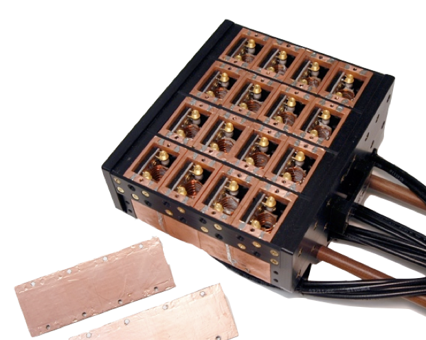
No, high throughput MRI is not an oxymoron.
The setup on their new Bruker system may be even more impressive, at least in terms of the financial investment. They have installed four CryoProbes (I think one costs around £200k!) in that one scanner for simultaneous multi-mouse in vivo imaging. Really cool, really expensive.
What’s a CryoProbe, you ask? It is a liquid helium-cooled RF surface array coil that can boost SNR by up to a factor of 7 compared to normal room-temperature coils. This allows them to acquire in vivo images at 60-micron isotropic resolution in under 2 hours. Wow. And remember, they have four of these that they run in parallel in one scanner.
There are trade-offs, of course. You can’t get the same level of per-animal optimisation in terms of things like shimming and controlling physiology, which are essential for the functional scanning we do at BRAIN. There’s also less flexibility – those CryoProbes are more or less always in the scanner; taking them out and putting them in is a hassle and (I believe) discouraged by Bruker – so you wouldn’t be able to scan mouse heads one day, mouse livers the next day, and rat heads the day after that (a somewhat regular occurrence at BRAIN). But if you just want to acquire a lot of high-resolution mouse brain structural scans, the MICe setup is king.

Low-res picture of four high-res mouse brain MR images acquired in parallel with four CryoProbes
Aside from a tour of the MICe lab and all of their fancy toys, the course covered a range of topics from basics of MRI to image registration to multimodal image analysis. Coming from cancer imaging, I didn’t really know much about neuroimaging. I had only heard of NIfTI, and I thought MINC was what old rich men bought their trophy wives. Yes, our peers in Canada don’t care much for NIfTI, and MINC is their data format of choice. They developed a library called RMINC for doing statistical analysis on MINC volumes in the R environment.
During the course, there were several hands-on tutorials where we got to use RMINC to analyse real data. That was what I liked best about the course – it was very practical, and as a neuroimaging novice, I got a lot out of it.
I am very grateful to the funders and everyone at MICe for putting together a great course. I think they’re doing it again next year, so if you have the opportunity to go, I highly recommend it.